 CLOSE SIDEBAR
Identification and histological mapping of senescent stromal cells in adipose tissue: a path towards tissue desenescence
Anthony S. Elias
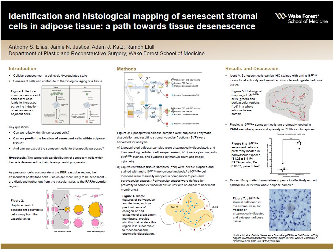
Background: Cellular senescence – a cell cycle dysregulated state – has widely been considered to be detrimental to tissue turnover and thus contributory to histotypical aging. Weeding out tissue resident senescent cells, hence “desenescing” aging tissues, may improve tissue upkeep while rendering senescent-rich cell populations. We hypothesize that the distribution of these cells within tissue is determined by their developmental progression: as precursor cells accumulate around the PERIvascular region, their descendant postmitotic cells – which are more likely to be senescent – are displaced further out from the vascular axles (the PARAvascular region). p16INK4a, a cyclin-dependent kinase inhibitor, is considered to be a cellular marker for senescence.
Hypothesis: By quantifying the population of p16INK4a-expressing cells in these regions, we aim to confirm the unequal distribution of senescent cells within adipose tissue.
Methods: Adipose whole tissue samples (n=5) were needle biopsied and stained with anti-p16INK4a monoclonal antibody. Equivalent samples were enzymatically dissociated, and their resulting isolated cell suspensions were cytospun, anti-p16INK4a stained, and quantified by manual count and image cytometry.
Results: p16INK4a+ stromal cells were readily present in enzymatically isolated cell suspensions: mononuclear bodies of 20-35 ø with a small nuclear/cytoplasm ratio, homochromatic nucleus, and clear cytoplasm. Initial manual cell count ranged between 1-3% in high power magnification fields, similar to preliminary image cytometry readings of approximately 4%. A number of large p16INK4a+ cluster structures were also identified; their cellular composition remains under study. In biopsied tissue samples, p16INK4a+ stromal cells were present with an approximate density of 70.33 ± 29.96 cells/mm3. Interestingly, p16INK4a+ cells were preferentially located in PARAvascular regions and sparsely in PERIvascular regions (81.23 ± 8.41% PARAvascular, P= 0.0007, paired t test).
Conclusions: Senescent stromal cells within adipose tissue are identifiable in lipoaspirated samples as p16INK4a+ cells. They are predominately housed within the paravascular regions, and enzymatic dissociation appears to effectively extract p16INK4a+ cells.
Source of mentor’s funding or other support that funded this research: Department of Plastic and Reconstructive Surgery, Wake Forest Baptist Health
Powered by Acadiate
© 2011-2024, Acadiate Inc. or its affiliates · Privacy