 CLOSE SIDEBAR
Effect of Weight Loss on Lumbar Bone Marrow Adipose Tissue in Older Males and Females
Quinn Powell
Poster Title: Effect of Weight Loss on Lumbar Bone Marrow Adipose Tissue in Older Males and Females
Student: Quinn Powell, Class of 2024
Faculty Mentor and Department: Dr. Ashley Weaver, PhD, Department of Biomedical Engineering
Funding Source: Clinical and Translational Science Institute of Wake Forest School of Medicine
Background: Older adults (65+ years) have an increased risk for age-related osteoporosis and fracture. This could be attributed to the loss of bone mineral density and increase in bone marrow adipose tissue (BMAT) seen in normal aging, as osteoblasts and adipocytes share a common stem cell progenitor line. BMAT infiltrates medullary cavity spaces that would otherwise be filled by bone, decreasing bone integrity. Bone marrow adipocytes display a different lipid metabolism from white adipocytes and increase in size during caloric restriction rather than decrease, meaning BMAT levels increase in response to typical weight loss (WL) interventions. This study aims to provide further data on BMAT variation during WL intervention in obese older adults, as well as evaluate for sex specific changes in BMAT with WL.
Hypotheses: (1) At baseline, participants with a higher BMI will have higher %BMAT in lumbar vertebrae. (2) After 6-months of weight loss, a greater decrease in BMI will be associated with a larger increase in %BMAT in the lumbar vertebrae. (3) There will be sex specific differences in BMAT associations when accounting for BMI.
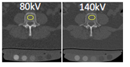
Methods: The study sample included 52 obese, older adults (27 females, 25 males). Using a Siemens SOMATOM Definition Flash dual source CT scanner, dual-energy (80 kV and 140 kV) helical CT scans were obtained of the L1-L5 vertebrae at baseline and after 6-months of weight loss. A Mindways Model 3 CT calibration phantom was positioned under each participant and imaged in each scan to calibrate BMAT measures. For each scan, a region of interest (ROI) was placed consistently in the trabecular region of each lumbar vertebra and in three adjacent mid-vertebral slices. These ROIs were used to derive basis material compositions and compute an average %BMAT for each lumbar vertebra for each participant. The three slices of each vertebra were then used to calculate an average %BMAT at each vertebral level. Both absolute and percent changes in %BMAT were used in analysis to account for baseline differences. Linear correlations were obtained to analyze relationships between baseline %BMAT vs BMI and Δ%BMAT vs ΔBMI for each lumbar vertebra (L1, L2, etc). Data were also stratified by sex.
Results: Higher baseline BMI was associated with lower %BMAT in all lumbar vertebrae [negative correlations, R = -0.55 (L1), -0.41 (L2), -0.61 (L3), -0.53 (L4) and -0.57 (L5)]. Greater increases in L2-L5 %BMAT were seen with more WL (ΔBMI) [negative correlations, R = -0.18 (L2), -0.22 (L3), -0.11 (L4) and -0.22 (L5)], although not at L1 [slight positive correlation, R = 0.02]. A Mixed Model analysis in JMP found no significant difference in percent change in %BMAT between the sexes (p = 0.84), nor a significant difference in %BMAT between lumbar vertebral level (p = 0.59).
Conclusions: Higher baseline BMI was associated with lower %BMAT. Larger increases in %BMAT were seen with greater WL (ΔBMI). No significant difference in Δ%BMAT was found between male and female participants.
Source of mentor’s funding or other support that funded this research: My role in analyzing and comparing BMAT levels in vivo is part of a larger ongoing clinical trial, UPLIFT: Utilizing Protein During Weight Loss to Impact Physical Function. UPLIFT funding source: K25AG058804 and R01AG050656
Powered by Acadiate
© 2011-2024, Acadiate Inc. or its affiliates · Privacy