 CLOSE SIDEBAR
Optimal anchor placement for collateral ligament reconstruction in elbow trauma- A 3D modeling study
Connor Dean
ABSTRACT
Background: Anchor fixation in the medial and lateral distal humeral columns is a critical step in many variations of MCL(UCL) and LCL repair in elbow trauma. The correct placement of surgical hardware in these procedures is critical for healing, recovery, and for avoiding revision surgeries. With the novel advancement of 3D modeling software there exists an opportunity to analyze anchor fixation with precision.
There is limited data that dives into specific dimensional analysis for ideal anchor placement in the distal humerus. Surgery to repair the LCL and MCL is technically challenging and revisions are common and may be related to failure of the implants due to suboptimal position of hardware or errors in bone tunnel placement in the distal humerus. 3D analysis is a novel approach to surgical planning allowing for an in-depth analysis of the proper positioning of hardware in the medial and lateral epicondyle / humeral columns with the aim of helping to prevent post-op complications and revisions.
Hypothesis: The hypothesis of this study is that 3D analysis of the medial and lateral epicondyles and columns of the distal humerus will allow us to define the optimal orientation and depth of anchors. “Optimal placement” is considered the position that provides the best implant fixation within the medial and/or lateral column of the distal humerus, while avoiding penetration 1) anteriorly into the coronoid fossa, 2) posteriorly into the olecranon fossa, or 3) far cortex from the point of insertion.
Methods: Subjects with CT scans of the distal humerus were reconstructed from DICOM stacks in Blender 3D modelling software using the OrthoMesh3D add-on module that was developed in our department. These models were analyzed for optimal anchor placement parameters into the medial and lateral epicondyles at the point of isometry. Data was collected relating to anchor depth, angle and position of when inserted into the medial and lateral columns of the distal humerus.
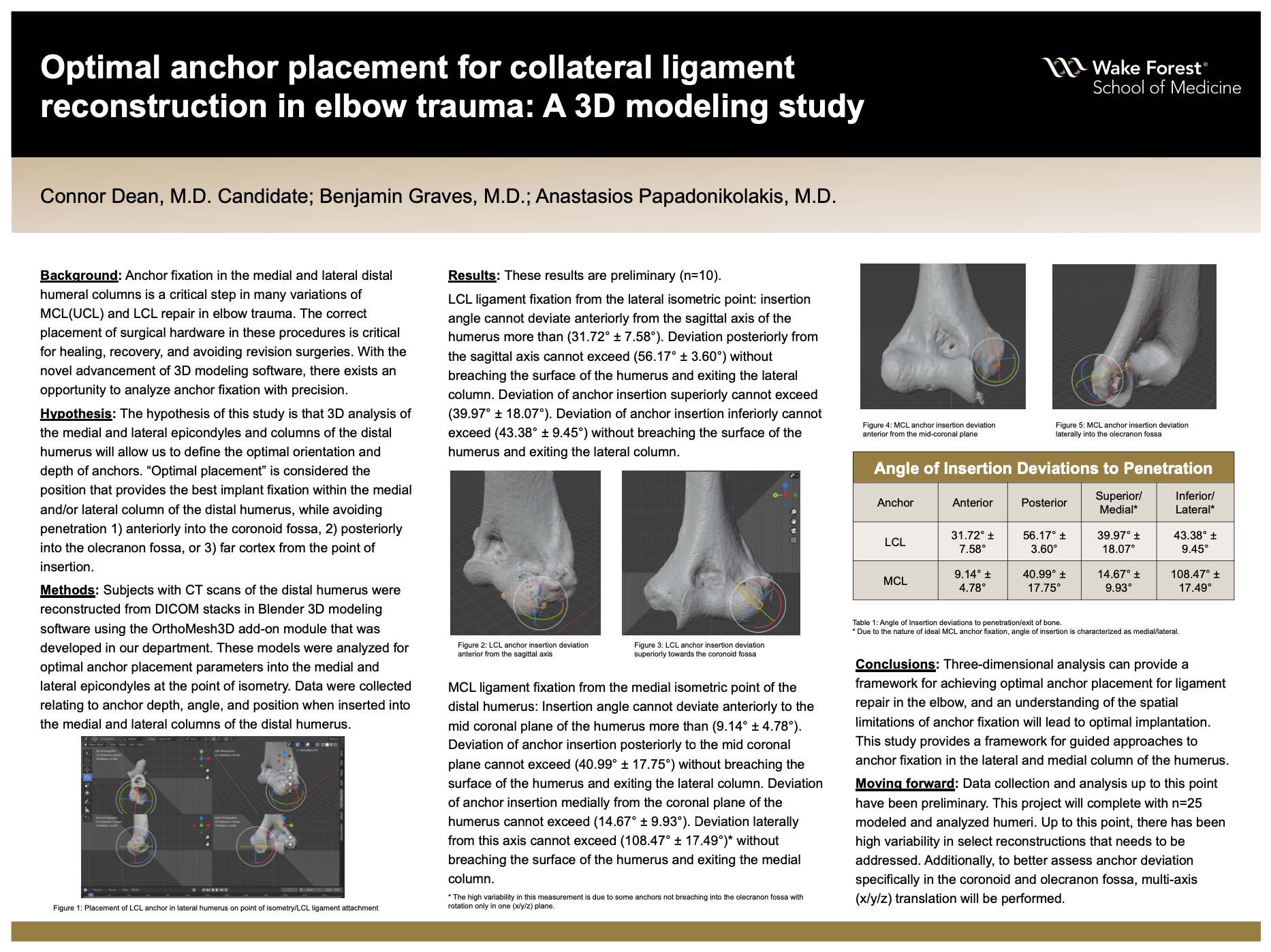
Results: These results are preliminary (n=10).
LCL ligament fixation from the lateral isometric point: insertion angle cannot deviate anteriorly from the sagittal axis of the humerus more than (31.72° ± 7.58°). Deviation posteriorly from the sagittal axis cannot exceed (56.17° ± 3.60°). without breaching the surface of the humerus and exiting the lateral column. Deviation of anchor insertion superiorly cannot exceed (39.97° ± 18.07°). Deviation of anchor insertion inferiorly cannot exceed (43.38° ± 9.45°) without breaching the surface of the humerus and exiting the lateral column.
MCL ligament fixation from the medial isometric point of the distal humerus: Insertion angle cannot deviate anteriorly to the mid coronal plane of the humerus more than (9.14° ± 4.78°). Deviation of anchor insertion posteriorly to the mid coronal plane cannot exceed (40.99° ± 17.75°). without breaching the surface of the humerus and exiting the lateral column. Deviation of anchor insertion medially from the coronal plane of the humerus cannot exceed (14.67° ± 9.93°). Deviation laterally from this axis cannot exceed (108.47° ± 17.49°). without breaching the surface of the humerus and exiting the medial column.
Conclusions: Three-dimensional analysis can provide a framework for approaching MCL and LCL ligament fixation in the elbow. An understanding of the spatial limitations of anchor fixation will lead to optimal implantation. This study provides a framework for guided approaches to anchor fixation in the lateral and medial column of the humerus.
Source of mentor’s funding or other support that funded this research: MSRP: Department of Student Affairs, Wake Forest School of Medicine
Powered by Acadiate
© 2011-2024, Acadiate Inc. or its affiliates · Privacy