 CLOSE SIDEBAR
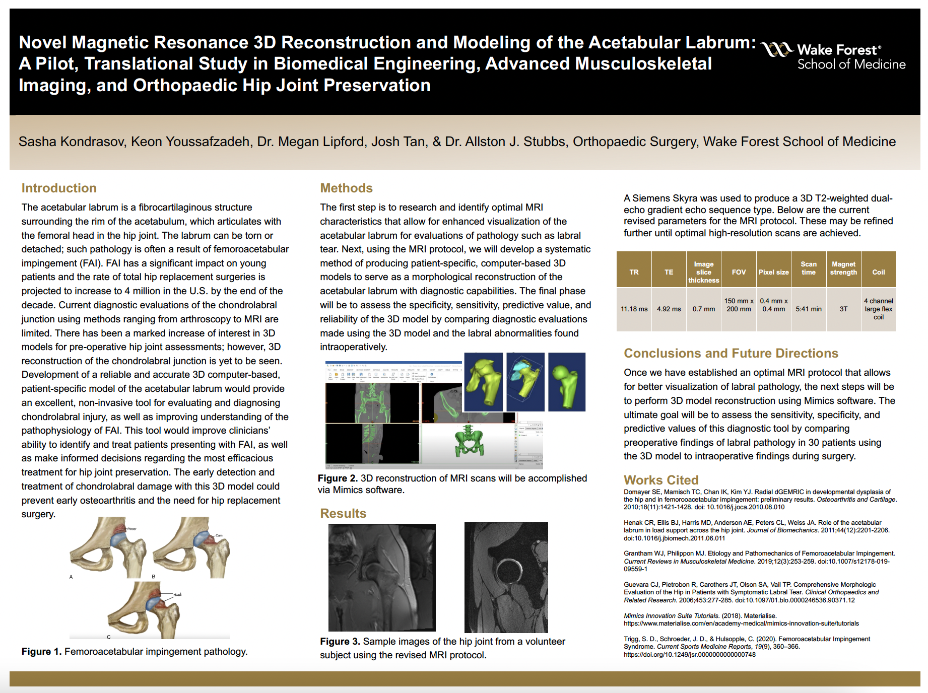
Novel Magnetic Resonance 3D Reconstruction and Modeling of the Acetabular Labrum: A Pilot, Translational Study in Biomedical Engineering, Advanced Musculoskeletal Imaging, and Orthopaedic Hip Joint Preservation
Sasha Kondrasov
Poster Title: Novel Magnetic Resonance 3D Reconstruction and Modeling of the Acetabular Labrum: A Pilot, Translational Study in Biomedical Engineering, Advanced Musculoskeletal Imaging, and Orthopaedic Hip Joint Preservation
Student: Sasha Kondrasov, Class of 2024
Faculty Mentor and Department: Dr. Allston Stubbs, Orthopaedic Surgery
Funding Source: Department of Medical Education, Wake Forest School of Medicine
ABSTRACT
Background: The acetabular labrum is a fibrocartilaginous structure surrounding the rim of the acetabulum, which articulates with the femoral head in the hip joint. The labrum can be torn or detached; such pathology is often a result of femoroacetabular impingement (FAI). FAI has a significant impact on young patients and the rate of total hip replacement surgeries is projected to increase to 4 million in the U.S. by the end of the decade. Current diagnostic evaluations of the chondrolabral junction using methods ranging from arthroscopy to MRI are limited. There has been a marked increase of interest in 3D models for pre-operative hip joint assessments; however, 3D reconstruction of the chondrolabral junction is yet to be seen. Development of a reliable and accurate 3D computer-based, patient-specific model of the acetabular labrum would provide an excellent, non-invasive tool for evaluating and diagnosing chondrolabral injury, as well as improving understanding of the pathophysiology of FAI. This tool would improve clinicians’ ability to identify and treat patients presenting with FAI, as well as make informed decisions regarding the most efficacious treatment for hip joint preservation. The early detection and treatment of chondrolabral damage with this 3D model could prevent early osteoarthritis and the need for hip replacement surgery.
Hypothesis: A patient-specific 3D computer-based model of the chondrolabral junction will allow clinicians to more effectively evaluate acetabular labral pathology prior to surgical interventions and will serve as an invaluable diagnostic tool with refined specificity, sensitivity, and reliability.
Methods: (1) Research and identify optimal MRI characteristics that allow for enhanced visualization of the acetabular chondrolabral junction for evaluations of pathology such as a labral tear. (2) Using the MRI protocol, develop a systematic method of producing patient-specific, computer-based 3D models to serve as a morphological reconstruction of the chondrolabrum with diagnostic capabilities. (3) Assess the specificity, sensitivity, predictive value, and reliability of the 3D model by comparing diagnostic evaluations made using the 3D model and the labral abnormalities found intraoperatively.
Results: A siemens Skyra was used to produce a 3D T2-weighted dual-echo gradient echo sequence type. The current revised MRI protocol is as follows: 11.18 ms TR, 4.92 ms TE, 0.7 mm image slice thickness, 150 mm x 200 mm FOV, 0.4 mm x 0.4 mm pixel size, 5:41 min scan time, 3T magnet strength, and 4 channel large flex coil. This is the first phase of the project; therefore, more results are pending.
Conclusions: It is our hope that with a patient-specific 3D model of the chondrolabral junction, clinicians will be able to more accurately and effectively assess the acetabular labrum in patients presenting with FAI and other hip joint injuries so that appropriate treatment can be determined. If the hypothesis is supported, potential next steps will be to evaluate whether this diagnostic tool can be applied to other osteoarthritic pathologies aside from those of the hip joint.
Source of mentor’s funding or other support that funded this research: N/A
Powered by Acadiate
© 2011-2024, Acadiate Inc. or its affiliates · Privacy